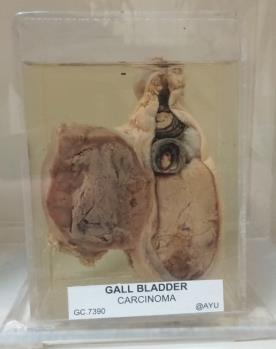
- Carcinoma of the Gallbladder
- GC.7390
- Histology Slide x2 Gallbladder laid open and showing in section a carcinoma of its wall. Excised from a female aged 70 years the subject of cholelithiasis. At the operation an irregular spherical mass projecting from one side of the gallbladder had contracted limited omental adhesions. The tumour and gallbladder were removed in one piece. On opening the gallbladder the fundus and the distal half were found packed with inspissated gallbladder contents. The cystic duct was blocked by a rounded gallstone and the space between it and the inspissated material was filled with two comparatively large facetted calculi and thirteen small facetted calculi. The gallbladder is slightly distended the fundus and adjacent half is filled with a greyish-white pultaceous-looking mass, and the rest of the cavity is filled with dark gallstones of moderate size. A gallstone of rather lighter colour is impacted at the commencement of the cystic duct. The gallbladder wall is not thickened and there is little evidence of pericholecystitis. The lateral wall of the gall bladder is indented by a spherical tumour some 4.3 cm in diameter. The surface of the tumour is nodular and part of it has the appearance of liver tissue. The tumour on section shows a marginal zone of grey homogeneous material around a more necrotic looking centre in which are a few small haemorrhages. Passing from the tumour towards the fundus of the gallbladder is a thin neoplasmic layer.
Height: 13.0 cm
Width: 11.5 cm
Depth: 8.2 cm
Length: Slide 7 cm
Width: Slide 2.5 cm


