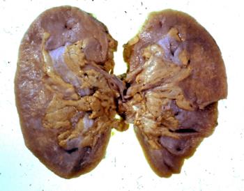
- Renal failure following uraemic ulceration of colon
- GC.11909
- Renal failure following uraemic ulceration of colon Clinical history: From a male aged 54 years. Admitted to hospital for investigation of headaches, with tiredness and lassitude. The patient was a known case of prostatic carcinoma, he was treated with oestrogens. Shortly after admission complained of lower abdominal pain of colicky character and began to pass diarrhoea with fresh blood. Was examined by sigmoidoscope, then a laparotomy was carried out, and the diagnosis of procto-colitis made. Following operation he never show improvement. he was known to be hypertensive, and retinae bore this out; renal function tests indicated very considerable impairment, and soon renal output shrank to nil, B.U.N. rose, and despite electrolyte imbalance being fairly adequately maintained he died. Post mortem findings: Genito-urinary system Both kidneys were reduced to less than half their normal bulk and each weighed 60 gms. Their capsules stripped readily to reveal a granular surface. On sectioning they showed marked reduction in cortical bulk and marked yellow pigmentation of tubules, presumably due to lipoid accumulation. The cortex was otherwise paler than normal and the appearances suggested an advanced degree of chronic nephritis. The calyces, pelvis, ureters were normal, the bladder was hypertrophied but was not inflamed and the prostate, which remained showed no macroscopic evidence of tumour. Alimentary system Tongue, pharynx: n.a.d. The oesophagus showed a mild degree of oesophagitis. The stomach and duodenum showed no change of note. The uppermost part the jejunum was rather congested and in many places in jejunum there were small, submucosal haemorrhages. In the mid portion of the jejunum several shallow. uraemic ulcers were present in the gut. Some portions of the ileal mucosa were congested but no ulcers were present in the ileum and in general the ileum looked much healthier than the jejunum. There was an abrupt change at the ileo-caecal valve however and the caecum showed very marked uraemic ulceration, which involved approximately 2/3 of the mucosa of the caecum. The ulcers were shallow and, were covered by a yellow-grey exudate. The mucosa of the appendix appeared normal. The gross uraemic ulceration continued for some distance into the ascending colon and from that point onwards to just passed the splenic flexure the mucosa was generally congested and there was a line of uraemic ulceration similar to that in the caecum along each taenia coli. No ulceration was detected in the pelvic colon or rectum and the mucosa appeared relatively healthy. The peritoneal cavity was normal. The liver weighed 1500g., was of normal shape, but on sectioning showed marked pallor due to anaemia and possibly also due to fatty change. Cardiovascular system: The pericardial sac was normal. The heart weighed 420 gms. and showed a moderate degree of left ventricular hypertrophy. The coronary arteries were rather atheromatous and one small focus about cm. diameter of fibrosis was detected in the left ventricular myocardium on the lateral walls The cardiac valves showed no significant abnormalities the other chambers were dilated There was a valvular patency of the foramen ovale and the upper portion of the septum secundum was fenestrated. Neither of these were likely to have caused any functional improvement during life. The aorta showed a relatively mild degree of atheroma, the pulmonary artery contained no thrombi.
- Twentieth century
Height: Jar 15 cm
Width: 15 cm
Depth: 6 cm
Weight: Kidney (each) 60 g


.jpg&width=350&height=350)