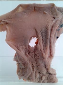
- Oesophagus showing instrumental rupture
- GC.14272
- Oesophagus showing instrumental rupture, from an 80 year old female. 6 months history of dysphagia. Barium swallow showed a constricying lesion at the junction of the middle and lower thirds of the oesophagus. On oesophagoscopya rigid smooth structure was present 35cm from the upper alveolus. biopsy showed cellular infiltration but no evidence of malignancy or fibrosis. A second biopsy and oesophagoecopy one month later showed epidermoid carcinoma. The stricture was dilated by oesophagoscopy and following this she developed a pain in the chest. X-ray did not disclose escape of air but gastrogafin demonstrated escape of content into the mediastinum. An emergency partial oesophagectomy was undertaken from which she made an uncomplicated recovery. The specimen is the section removed, and shows in the lower part the area of constriction where the mucosa is raised and forms a series of nodules projecting into the lumen but there is no gross evidence of ulceration. Proximal to this the oesophagus is dilated and smooth and the punched out hole with somewhat ragged edges is present immediately above the site of the tumour. This has passed through all coats of the oesophagus Perforation of the wall of the viscus during oesophagoscopy is a hazard when rigid instruments are used for this purpose. it is much less likely to happen when using malleable fibre glass instruments. Histological description in gc ledger
- Twentieth century
Height: container 11.3 cm


